Scientists have shown what the Covid-19 coronavirus looks like under a microscope
Categories: Health and Medicine | Microworld | Science | World
By Pictolic https://pictolic.com/article/scientists-have-shown-what-the-covid-19-coronavirus-looks-like-under-a-microscope.htmlOn Thursday, February 13, the National Institute of Allergy and Infectious Diseases showed the first images of the Covid-19 virus (also informally known as 2019-nCoV or SARS-CoV-2), which affected more than 60,000 and killed 1,370 more people. Since its manifestation in December last year, almost all media outlets have been constantly trumpeting about it.

Viruses are tiny objects consisting of DNA or RNA wrapped in a protein layer. They are too small to be seen with a conventional light microscope.

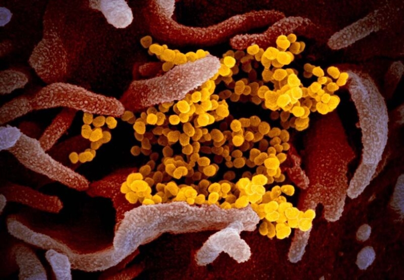
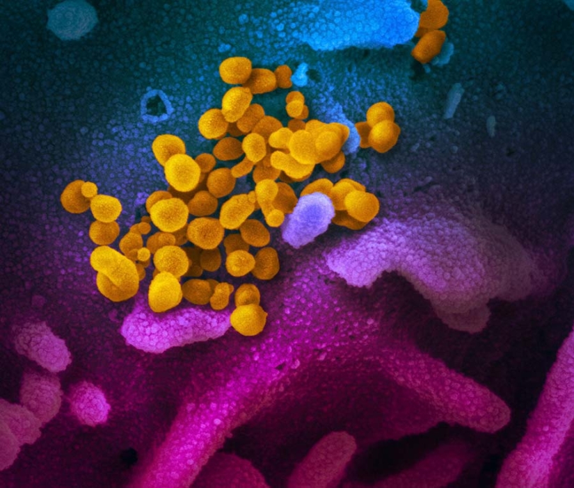
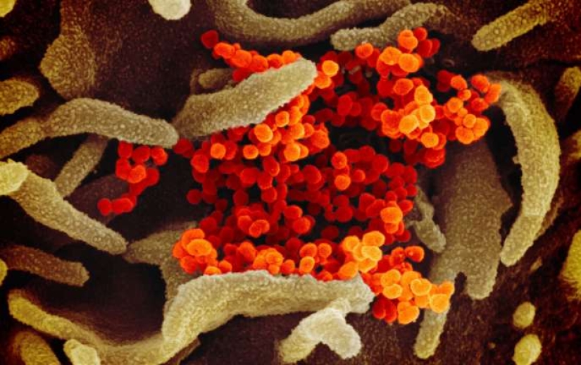
Scientists have posted online images of the coronavirus obtained using scanning and transmission electron microscopes — and, oddly enough, viruses surprisingly look aesthetically pleasing.

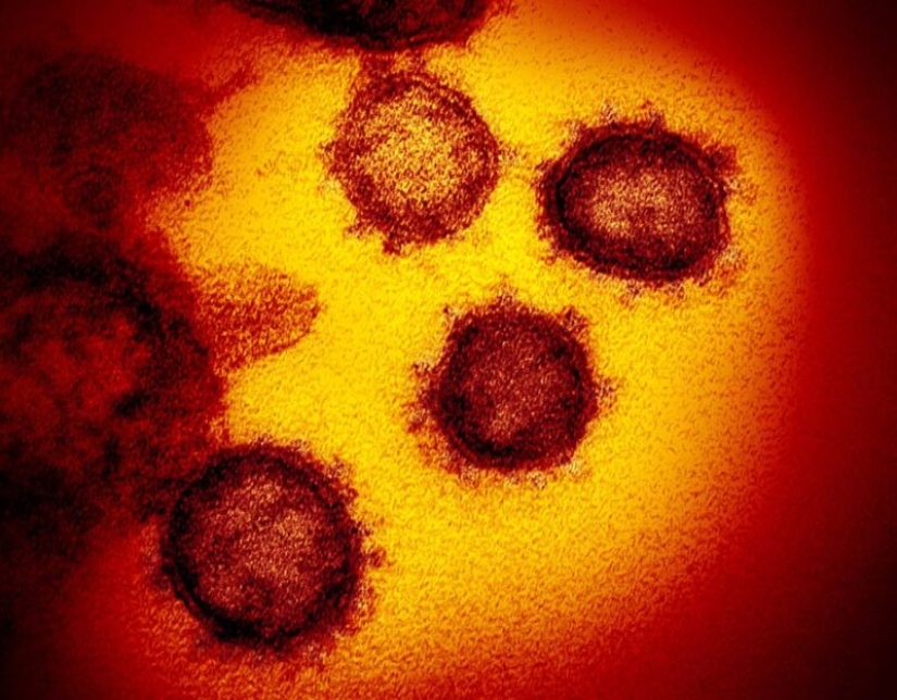
The image above was obtained using a transmission electron microscope. It's not as sharp as the first one, but you can see the spikes on the surface of the virus, thanks to which the coronavirus got its name. If the pictures look familiar, it's because most coronaviruses, for example, such as SARS and MERS, look almost the same externally.Viruses in the coronavirus family have only small differences in their genome, and there are only 5 nucleotide differences between 3 viruses. However, viruses can have markedly different manifestations when it comes to infecting people.

The images you see in this material are the result of teamwork. RML researcher Emmie de Wit provided the virus, microscopist Elizabeth Fischer created the images, and the Department of Visual Medical Art colored them.
Keywords: Coronavirus | Microscope | Microphoto
Post News ArticleRecent articles

All cultures, countries and different Nations, each nation has its own traditions and customs. But one thing remains common — ...

They say that absolutely everything that is made of chocolate is delicious. And apparently, Sarah Hardy agrees with this opinion. ...
Related articles

In this difficult time of quarantine and pandemic many people deal with boredom and stress, sitting alone in four walls and not ...

It may seem that nothing can't be hidden, but also the most goggle-eyed of us are unlikely to consider a speck of dust. In such ...

As in most countries of the world, when a child is born in a Japanese family, relatives come to visit with gifts. Due to the ...

New Jersey photographer Robin Schwartz photographed her adorable daughter Amelia with a variety of animals from 2002 to 2015. The ...