Paper Anatomy
Categories: Exhibition | Health and Medicine
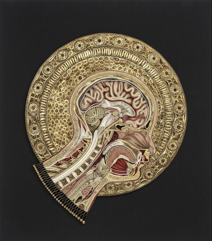
By Pictolic https://pictolic.com/article/paper-anatomy.htmlThese human organs are made from Japanese paper and the gilded edges of old books using the quilling technique. Quilling first appeared in the Renaissance era — it was invented by monks and nuns who found a way to use the gilded edges of old bibles, and later — by the XVIII century — aristocratic ladies created masterpieces in the quilling technique in their spare time.
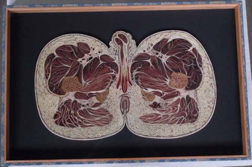
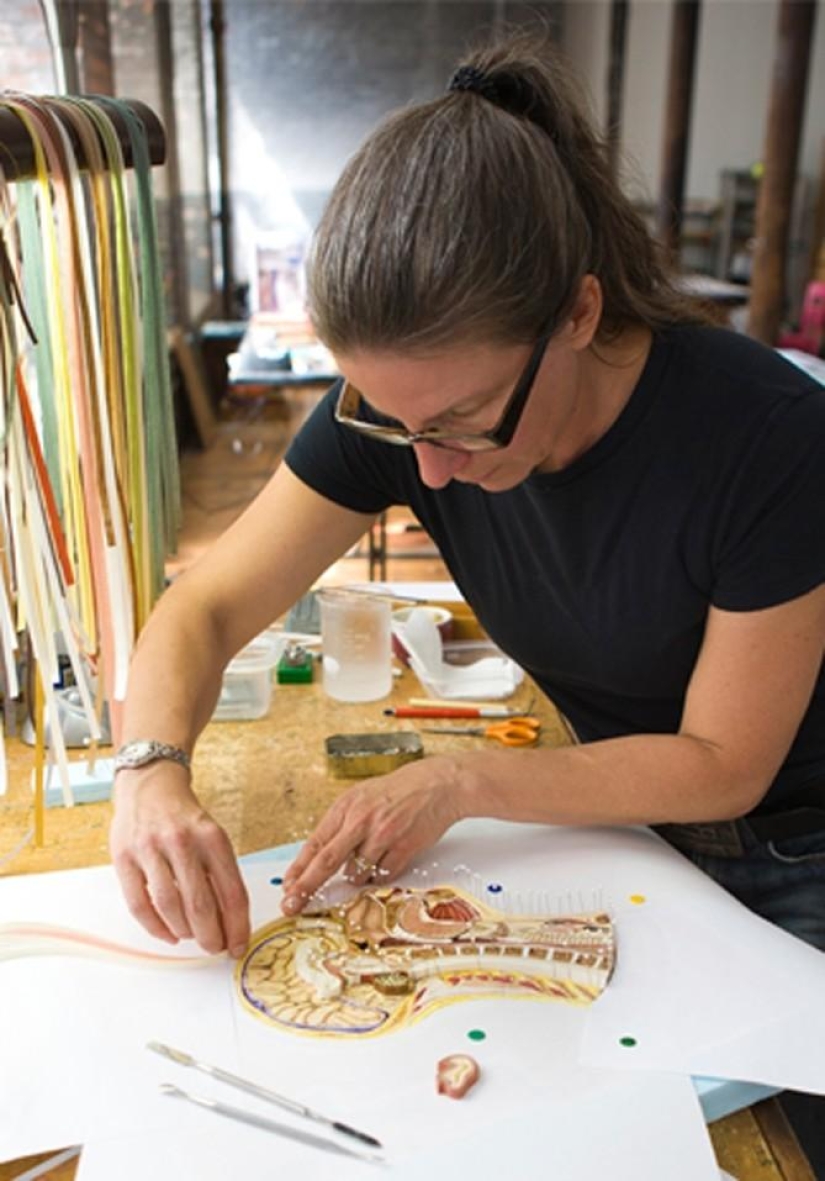
Artist Lisa Nilsson has found an interesting way to use various forms of paper folded in the quilling technique — she creates mind-blowing models of human organs.
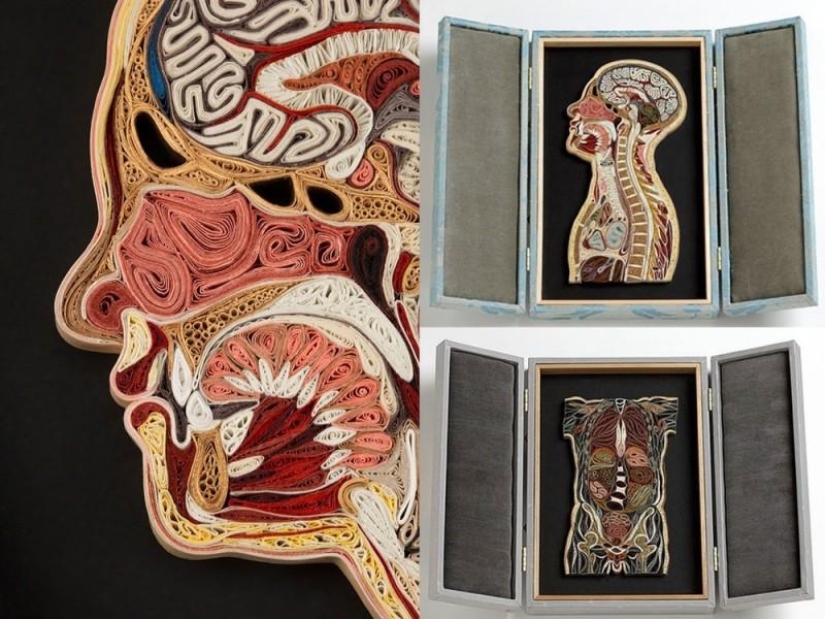
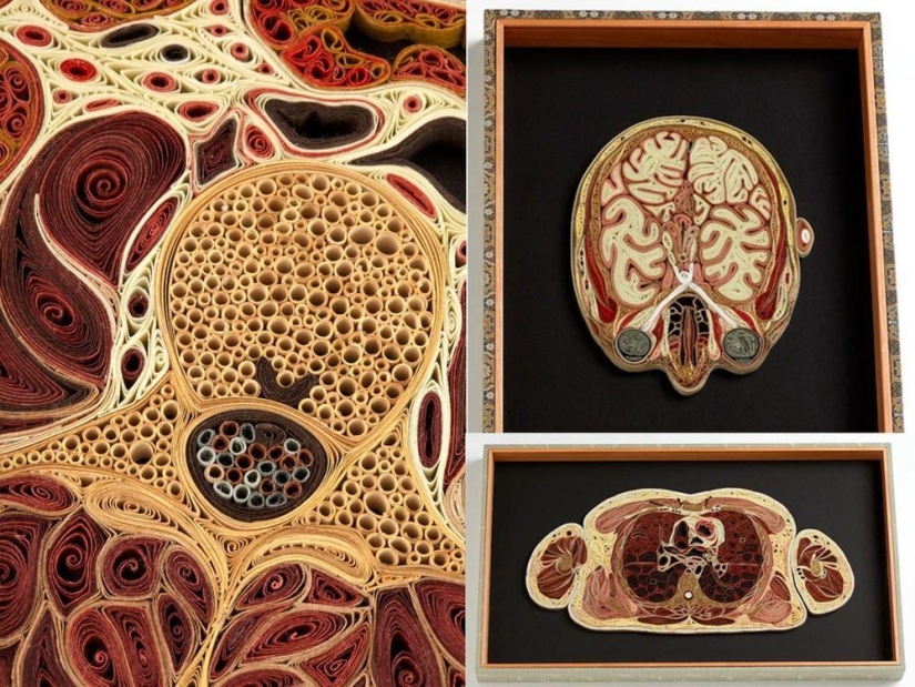
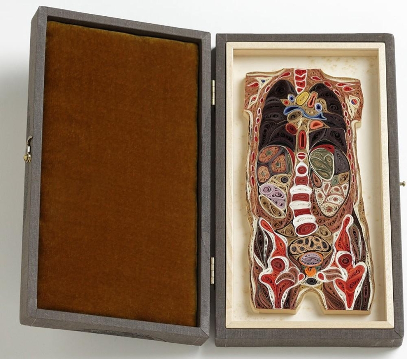
Lisa Nilsson, a graduate of the Rhode Island School of Design, created the Tissue project. This is a paper image of the anatomy of the human body. The works are made in the quilling technique, made of Japanese mulberry paper.
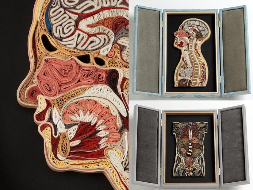
It takes from several weeks to several months to create each such model from twisted strips of paper. The work begins with Lisa pinning strips of paper on photos of real cuts, and gradually fills the space with missing details.
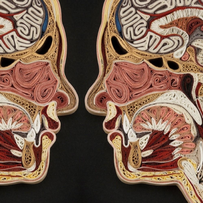
From the outside, all these internal organs in the section, the structure of the skull, skeleton and other anatomical images seem to be painted or molded from plaster. But if you look closely, it becomes clear — every bone, every vein, vein and muscle of the anatomical exhibits are made of multicolored, neatly twisted strips of paper.
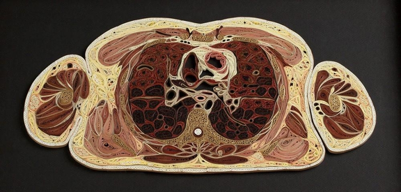
Each of her creations turns out to be colorful and natural. And although some anatomical sections of humans and animals are shocking, it is impossible not to admire Lisa's meticulous work.
Recent articles

It's high time to admit that this whole hipster idea has gone too far. The concept has become so popular that even restaurants have ...

There is a perception that people only use 10% of their brain potential. But the heroes of our review, apparently, found a way to ...

New Year's is a time to surprise and delight loved ones not only with gifts but also with a unique presentation of the holiday ...